SOIL TRANSMITTED HELMINTHS
SOIL TRANSMITTED HELMINTHS
INTRODUCTION
Soil-transmitted helminth (STH) infection is highly endemic in tropical and subtropical areas of sub-Saharan Africa, Asia and Latin America, where up to 2 billion people have active infections. STH disease has mainly remained neglected by the global health community because the people most affected are among the most impoverished and because the disease causes chronic ill health with insidious clinical presentations, rather than severe acute illness or high mortality.However, it is now recognised that STH infection causes significant morbidity worldwide with 39 million disability-adjusted life years (DALYs) lost each year - more than those lost to malaria (36 million yearly) and approaching those lost to tuberculosis (47 million annually). Hookworm infection alone causes
a loss of 22 million DALYs.
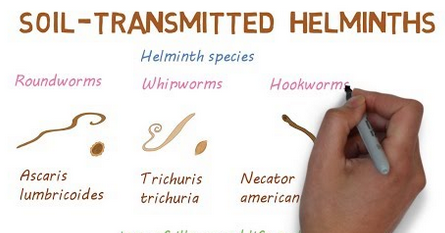
Ascaris lumbricoides (large roundworm of man) Infection with this roundworm is widespread, with estimates of the annual incidence of infection being higher than 1500 million cases, or around one quarter of the world's population. In addition to the species in man, Ascaris lumbricoides, a morphologically indistinguishable species Ascaris suum is found in the pig. Other related genera include Parascaris in equines and Toxascaris in a variety of domesticated animals.
Morphology
The adult Ascaris lumbricoides are largely white, or pinkish-white, cylindrical roundworms, slightly narrower at the head. The more slender male's measure between 10 to 30cm long and have a curved tail with two spicules, but no copulatory bursa. The females are very similar, being slightly larger at between 20 to 35cm long, a vulva approximately a third of the length of the body down fromthe head, and have a blunt tail. They are both characterised by having a smooth, finely striated, cuticle, and a mouth, which is characteristic of all of the Ascarids (e.g. Toxocara), having three lips each equipped with small papillae. Internally they follow the generalised body plan of all nematodes and have a cylindrical oesophagus opening into a flat ribbon-like the intestine. The eggs consist of a thick transparent inner shell which is covered in a thick, warty, albuminous coat.
Life cycle
These parasites have a direct life cycle, with no intermediate hosts. The adult parasite lives in the lumen of the small intestine of man, usually only feeding on the semi-digested contents of the gut, although there is some evidence that theycan bite the intestinal mucous membrane and feed on blood and tissue fluids. The female parasite is highly prolific, laying an estimated 2 million eggs daily. In the intestine, these only contain an unembryonated mass of cells, differentiation occurring outside the host.This requires a temperature less than 30°C, moisture and oxygen, before the development of the young L1 larvae after approximately 14 days. Eggs containing the L2 larvae take another week to develop, before they are infective to man, and may remain viable in the soil for many years if conditions are optimal. Infection occurs on ingestion of raw food, such as fruit or vegetables, that is contaminated with these infective eggs. The eggs then hatch in the small intestine, to release the L2 rhabditiform larvae (measuring approximately 250 by 15µm in size.
These do not only grow into the adult forms in the intestine, but must then undergo a migration through the body of their host. These L2 larvae penetrate the intestinal wall, entering the portal blood stream, and then migrate to the liver, then heart, then after between 1 to 7 days, the lungs. Here they moult twice on the way to form the L4 larvae, (measuring approximately 1.5mm Long), then burrow out of the blood vessels, entering the bronchioles. From here they migrate up through the air passages of the lungs, to the trachea. They then enter the throat and are swallowed, finally ending up in the small intestine where they mature and mate, to complete their life cycle
Pathology of Infection.
The majority of infections (~85%) appear to be asymptomatic, in that there is no gross pathology seen. However, the presence of these parasites seems to be associated with the same general failure to thrive in their hosts seen with many of these intestinal nematodes. In terms of more easily identified pathology, this may be divided into three areas; Pathology Associated with the Ingestion and Migration of Larvae Severe symptoms of Ascaris infection may be associated with the migratinglarvae, particularly in the lungs.
If large numbers of these larvae are migrating through the lungs simultaneously, this may give rise to severe haemorrhagic pneumonia. More commonly, as is the case with most infections, the haemorrhages are smaller in scale, but still may lead to breathing difficulties, pneumonia and/or fever.
A complication here is that many of the parasites proteins are highly allergenic. Due to this, the presence of the migrating larvae in the lungs is often associated with allergic hypersensitivity reactions such as asthmatic attacks, pulmonary infiltration and urticaria and oedema of the lips. Pathology Associated with Adult Parasites in the Intestine The most common symptoms of infection are due to the adult parasite, and consist of rather generalised digestive disorders, such as a vague abdominal discomfort, nausea, colic (e.t.c.).
These symptoms are dependent to some extent on the parasite’s burden of the host, which in severe cases may consist of many hundreds or even thousands of parasites, although these are extreme cases. In the case of
these heavy infections, the presence of many of these large parasites may contribute to malnutrition in the host, especially if the hosts (often children) are undernourished.
A more serious, and potentially fatal, the condition may arise in these more heavy infections, where the mass of worms may block the intestine and need to be surgically removed. This may also occur sometimes on treatment for other. intestinal nematodes such as hookworms, where the curative drug dose for these parasites irritate the ascarids. Pathology due to "Wandering" Adults outside of the Intestine Adult parasites often leave the small intestine to enter other organs, (sometimes in
response to anti-helminthic drugs used to treat other intestinal nematode infections), where they may cause various types of pathology, sometimes with severe consequences.
For example, adult Ascaris worms may migrate to the bile duct, which may then become blocked, causing jaundice and general interference in fat metabolism. Adult parasites may also migrate to the appendix, or through the intestinal wall, both conditions which may cause fatal peritonitis as they may well carry intestinal bacteria to these sites. They may, alarmingly, sometimes migrate forward through the intestinal tract, to be either vomited up or emerging through the nose. More seriously, if they enter the trachea, they may cause suffocation.
Diagnosis
Definitive diagnosis is by the demonstration of the characteristic eggs in faecal samples or by identifying adult worms passed out spontaneously by the host.Epidemiology and Control
Infection occurs through ingestion of parasites’ eggs in the food. The eggs are highly resistant to adverse environmental conditions. This with other factors highlighted below are often associated with the transmission of infection;- Lack of or inadequate waste disposal facilities
- Improper washing of hands before eating
- Incorrect washing of fruits and vegetables before consumptions
- Unkept rooms and dwelling places that harbour mechanical carriers of parasites, etc.
Provision of the good waste disposal system and good personal hygiene will help to control infections.
0 comments